|
A 46 year-old woman presented with a left hemiplegia and headache. The medical history was significant for hypertension. |
![]()
![]()
![]()
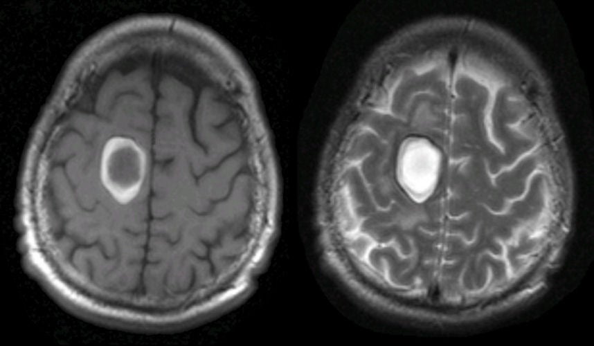
| Lobar Intracerebral Hemorrhage:
(Left) T1-weighted axial MRI; (Right) T2-weighted axial MRI. Note the large hemorrhage in the right frontal lobe.
Blood has
a complex pattern on MRI, depending on whether the image is T1- or T2-weighted, and how
long the hemorrhage has been present. The bright signal on T1-weighted and T2-weighted
images represents extracelluar methemoglobin. By contrast,
intracellular methemoglobin is bright on T1-weighted but dark on
T2-weighted images. If one looks closely,
one can also see a thin black surrounding
rim on T1-weighted and T2-weighted images. This is hemosiderin. These findings mark the hemorrhage as subacute. The classic locations for hypertensive intracerebral hemorrhages are the basal ganglia, thalamus, pons and cerebellum. However, hypertension can also result in lobar hemorrhages. In these cases, it is essential to exclude other causes of bleeding, including an underlying vascular malformation or tumor. In the very elderly, amyloid angiopathy is a common cause of lobar hemorrhage. The symptoms depend on the location of the hemorrhage. |
Revised
11/30/06.
Copyrighted 2006. David C Preston