|
A 55 year-old man presented with a headache and receptive aphasia. The medical history was significant for hypertension. |
![]()
![]()
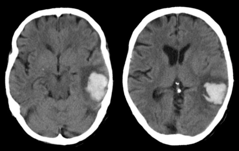
| Lobar Intracerebral Hemorrhage:
Axial CT scans. Note the large hemorrhage in the left
temporal lobe with surrounding edema. The classic locations for hypertensive intracerebral hemorrhages are the basal ganglia, thalamus, pons and cerebellum. However, hypertension can also result in lobar hemorrhages. In these cases, it is essential to exclude other causes of bleeding, including an underlying vascular malformation or tumor. In the very elderly, amyloid angiopathy is a common cause of lobar hemorrhage. The symptoms depend on the location of the hemorrhage. |
Revised
11/23/06.
Copyrighted 2006. David C Preston