|
A 72 year-old man noted chronic low back pain, worse with standing and walking. With prolonged standing, he would develop pain and numbness radiating down the posterior thighs and calves. |
![]()
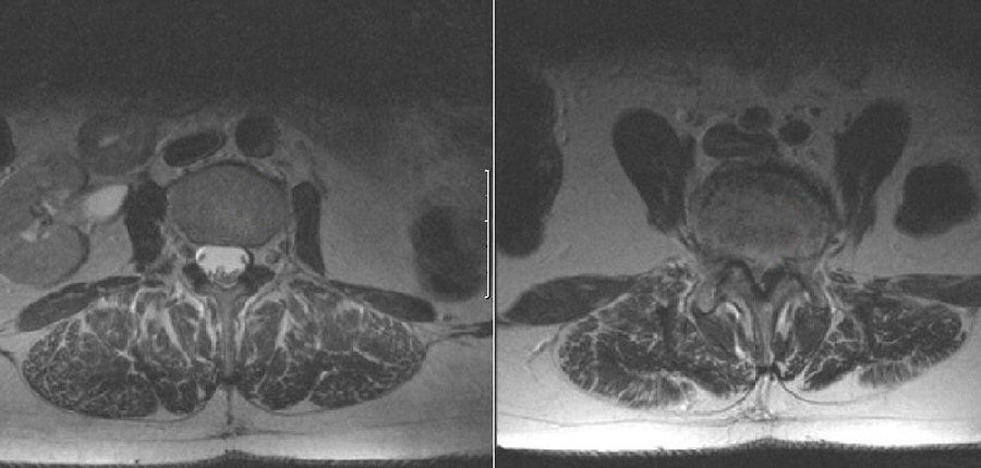
| Lumbar Spondylolithesis:
(Left) T2-weighted axial MRI at the
level of L3; (Right) T2-weighted axial MRI at the L4-5 disk level.
Note the severe central canal stenosis at the L4-L5 disk level
(right scan), with the complete absence of CSF (which is bright) and
compression of the nerve roots at this level. In contrast, at the L3
level (left scan) one can clearly visualize the CSF surrounding the
descending nerve roots of the cauda equina within the thecal sac.
Spondylolisthesis is defined as slippage (either forwards or backwards) of one vertebral body over the one beneath it. Spondylolisthesis may be asymptomatic or may cause back pain, radicular pain, or neurogenic claudication. It may be caused by a congenital abnormality, but more often results from a stress fracture or degenerative changes in the pars interarticularis (the area of bone at the point where the pedicle meets the lamina, articular facets and transverse process). |
Revised
11/30/06
Copyrighted 2006. David C Preston