|
A 72 year-old woman presented with progressive pain, weakness and sensory loss in the left leg. Her examination showed weakness of ankle and toe dorsiflexion as well as ankle eversion on the left. There was also loss of sensation over the lateral calf and dorsum of the foot. |
![]()
![]()
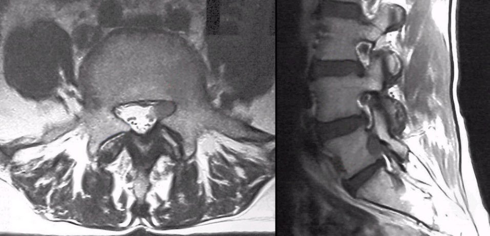
| Neurolymphomatosis:
(Left) T2-weighted axial MRI at the L5
level; (Right) T1-weighted
sagittal MRI. On the left scan, note the markedly
enlarged nerve root compared to the normal size of the other nerve
roots in the cauda equina (CSF is bright white, nerve roots are dark). On the right scan, the normal exiting
nerve roots can be seen surrounded by epidural fat. However, the
nerve root exiting below the L5 pedicle is markedly enlarged, filling the
entire foramen. Biopsy demonstrated infiltration of the nerve roots by malignant
lymphoma. It is important to remember that radiculopathy can be caused not only by
external compression (e.g., disk, spondylosis) but also by
infiltration of the nerve root by tumor or granulomatous tissue. Neurolymphomatosis may occur in isolation, or as part of widely systemic lymphoma either during the initial diagnosis or as the first sign of relapse in a treated patient. It is much more frequently seen in non-Hodgkins compared with Hodgkins lymphoma. It presents as a progressive sensorimotor polyneuropathy, cauda equina syndrome or isolated mononeuropathy (the sciatic nerve being the most common). |
Revised
11/25/06
Copyrighted 12007. David C Preston