 |
|
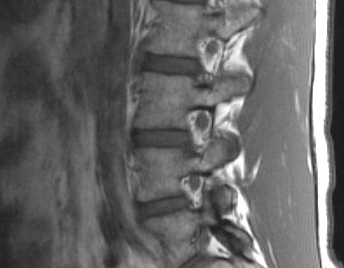
| Parasagittal MRI of the Lumbar Spine - T1-weighted image. Note that this slice provides an unique view of the exiting nerve roots through the intervertebral foramina. The nerve roots exit directly below the pedicles of the corresponding levels. As fat is bright on T1, the exiting nerve roots are well seen, being surrounded by epidural fat. |