 |
|
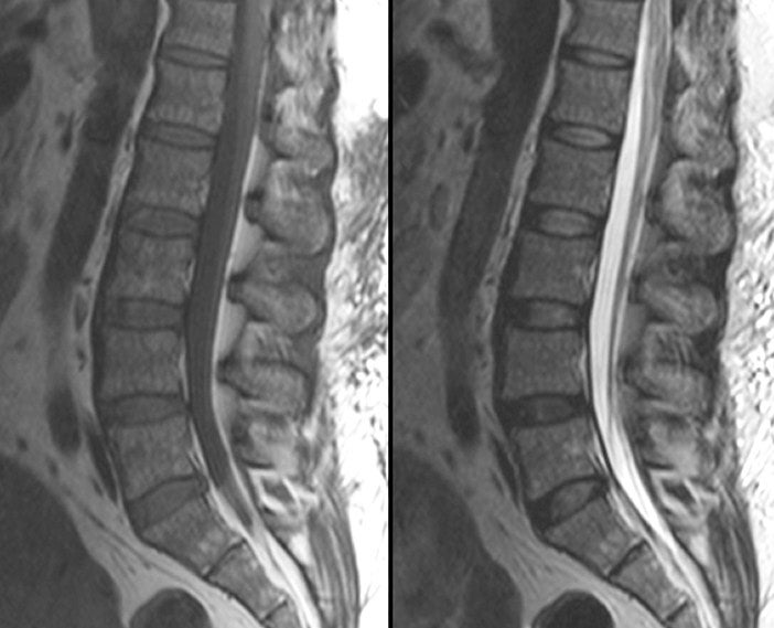
| Mid-Sagittal MRI of the Lumbar Spine. Left (T1-weighted image); Right (T2-weighted image). On this view centered over the lumbar spine, one can see all five lumbar vertebrae in addition to the sacrum and lower thoracic vertebrae. Note on the T1 image that CSF is dark and on the T2 image that CSF is bright. Also note the subcutaneous fat which is bright on both T1- and T2-weighted images. The mid-sagittal view is the optimal view to visualize the spinal cord, cauda equina, CSF and vertebral bones. However, as most disk herniations are posterior lateral (as opposed to straight posterior), the mid-sagittal view may not the optimal view to see a disk herniation. |