|
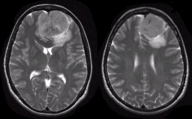
A 55 year-old woman presented with a change in personality. On exam, she was markedly abulic. |
![]()
![]()
![]()
| Meningioma (Parafalcine). T2-weighted
axial MRIs. Note the large mass that is dural based and compresses the adjacent frontal lobe and genu
of the corpus callosum. The mass is in the midline, arising from the left. This is the typical appearance of a meningioma. Meningiomas are common tumors that arise from the meninges and can occur within the spinal canal as well as intracranially. They are typically benign histologically, and can be surgically resected if they are in an accessible location. They are more common in women than men, and account for approximately 20% of all primary brain tumors. Similar to low grade gliomas, they grow very slowly. They may result in seizures, focal neurological signs, or both, depending on their location. Some of the more common locations for meningiomas include: ● Parasagittal (attached to the falx) |
Revised
11/30/06.
Copyrighted 2006. David C Preston