|
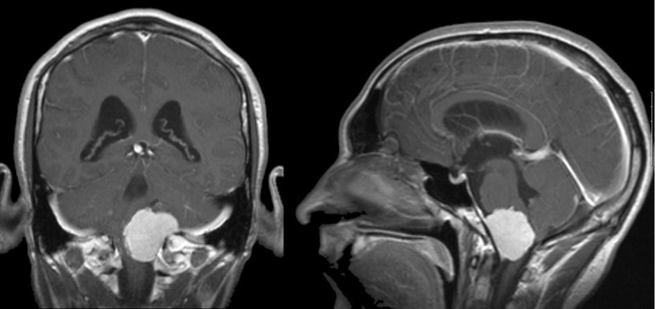
A 52 year-old woman presented with headaches, vertigo and falling. |
![]()
| Meningioma (Clivus). (Left) T1-weighted
with gadolinium coronal MRI; (Right) T1-weighted with gadolinium
sagittal MRI. Note the well demarcated mass that
enhances with gadolinium, and nearly obliterates the medulla. On the sagittal
scan, one can more easily see that the mass is dural based and arises from the clivus. Also note
that the cerebral aqueduct is enlarged, reflecting the
underlying obstructive hydrocephalus. Meningiomas are common tumors that arise from the meninges and can occur within the spinal canal as well as intracranially. They are typically benign histologically, and can be surgically resected if they are in an accessible location. They are more common in women than men, and account for approximately 20% of all primary brain tumors. Similar to low grade gliomas, they grow very slowly. They may result in seizures, focal neurological signs, or both, depending on their location. Some of the more common locations for meningiomas include: ● Parasagittal (attached to the falx) |
Revised
11/26/06.
Copyrighted 2006. David C Preston