|
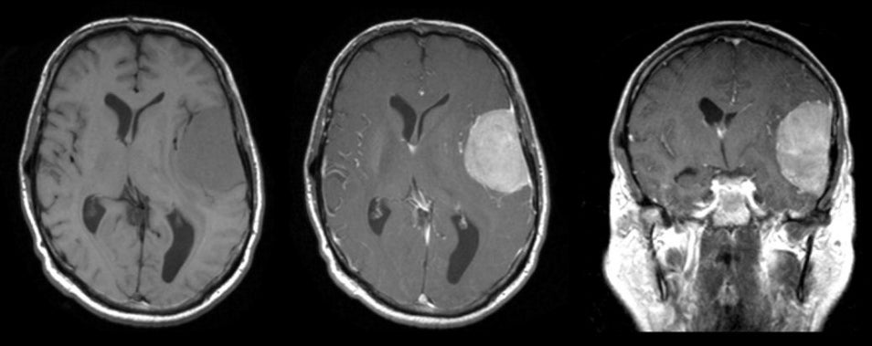
A 61 year-old woman presented with a three month history of receptive and expressive aphasia. On exam, there was a subtle right hemiparesis. |
![]()
![]()
![]()
![]()
| Meningioma (Frontal Operculum). (Left)
T1-weighted axial MRI; (Middle) T1-weighted with gadolinium axial MRI; (Right) T1-weighted
with gadolinium coronal MRI. Note the large, well demarcated mass
that is dural based and
compresses the adjacent frontal operculum. The mass directly
compresses the primary language area. It is isointense on T1-weighted
images but strongly
enhances with contrast. This is the typical appearance of a meningioma.
Meningiomas are common tumors that arise from the meninges and can occur within the spinal canal as well as intracranially. They are typically benign histologically, and can be surgically resected if they are in an accessible location. They are more common in women than men, and account for approximately 20% of all primary brain tumors. Similar to low grade gliomas, they grow very slowly. They may result in seizures, focal neurological signs, or both, depending on their location. Some of the more common locations for meningiomas include: ● Parasagittal (attached to the falx) |
Revised
11/30/06.
Copyrighted 2006. David C Preston