|
A 53 year-old woman presented with headaches. Her exam was normal. |
![]()
![]()
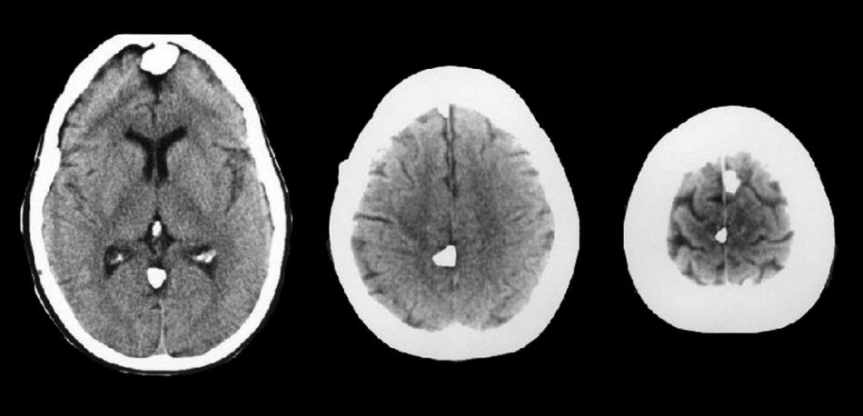
| Multiple Meningiomas. Axial
CT scans; (Left)
level of the basal ganglia; (Middle) level of the centrum semiovale; (Right)
level of the vertex. Note the multiple calcified meningiomas, all of which arise
from the falx. These are the typical appearances and locations of meningiomas. Meningiomas often calcify and are well seen on CT scan.
Meningiomas are common tumors that arise from the meninges and can occur within the spinal canal as well as intracranially. They are typically benign histologically, and can be surgically resected if they are in an accessible location. They are more common in women than men, and account for approximately 20% of all primary brain tumors. Similar to low grade gliomas, they grow very slowly. They may result in seizures, focal neurological signs, or both, depending on their location. Some of the more common locations for meningiomas include: ● Parasagittal (attached to the falx) |
Revised
11/30/06.
Copyrighted 2006. David C Preston