|
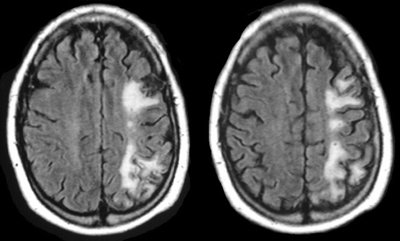
A 62 year-old man had an upper GI bleed complicated by severe hypotension. Several days after successful surgical repair of a bleeding gastric ulcer, he was noted to have significant weakness of the right shoulder and hip girdle muscles. |
![]()
| Watershed Infarction:
Flair axial MRIs: Note the linear area of infarction between the
distributions of the middle cerebral artery (MCA) and anterior cerebral artery
(ACA) on the
left side of the brain.
The area between two vascular territories is known as a watershed. Watershed infarcts typically occur following reduced perfusion pressure, often secondary to cardiac events or severe bleeding. In those cases, they are usually bilateral. When a watershed infarct is seen unilaterally, this is usually due to hemodynamic narrowing of a proximal artery (in this case, the carotid artery) without intact collateral vessels (e.g., an incomplete circle of Willis), with or without superimposed hypotension. Most commonly, these involve the distal territory of the carotid artery. Thus, if a patient has severe, hemodynamically significant carotid stenosis, then ischemia first occurs between the terminal territories of the middle and anterior cerebral arteries on that side. The watershed territory between the MCA and ACA corresponds to the shoulder and hip girdle muscles on the motor homunculus, leading to a characteristic clinical deficit, weakness of the shoulder and hip girdle muscles bilaterally (often referred to as "the man in the barrel" distribution of weakness). There is also a watershed territory between the MCA and PCA. When an infarct occurs in this territory, patients typically develop bilateral cortical visual abnormalities, among them cortical blindness, Anton's syndrome (cortical blindness with denial/confabulation) and Balint's syndrome (asimultagnosia, optic ataxia, and gaze apraxia). Lastly, a watershed area exists between deep and superior cortical vessels. Most often, this is in the basal ganglia / internal capsule areas which are supplied by the lenticulostriates below and the cortical branches above. |
Revised
10/12/06
Copyrighted 2006. David C Preston