|
An 82 year-old man became confused after a fall. Within a few days, he developed a language disturbance and mild right hemiplegia. The next day, he lapsed into a coma and displayed decerebrate posturing. |
![]()
![]()
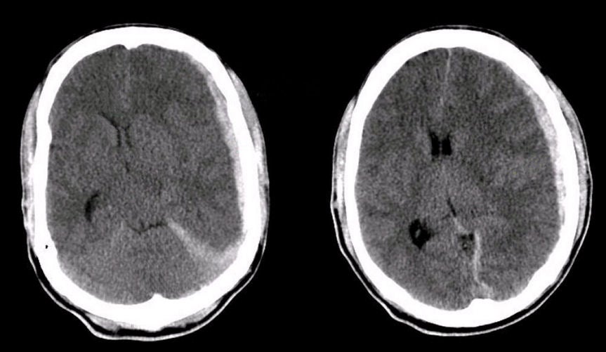
| Midline Shift and Impending Herniation:
Axial
CT scans of the brain at the level of the basal ganglia. Note
the sudural hematoma (SDH) on the left extending down to the tentorium (the flap of
dura that separates the cerebral hemispheres from the posterior
fossa). There is a resulting midline shift. When a
line is drawn down the midline, the amount of horizontal shift can be
measured. In this case, there is a 10.8 mm shift of midline (note the
septum pellucidum that separates the lateral ventricles should be in the
midline). This amount of shift is usually associated with coma.
The displacement of brain structures described above illustrates the Monro-Kellie doctrine, which states that in an adult the cranial volume is a constant. The cranial contents consist primarily of brain, cerebrospinal fluid (CSF) and blood vessels. If a mass such as a hematoma, tumor or edema develops, these elements must shift to accommodate the mass. Since the cranial volume is a constant, part of the cranial contents will herniate through the tentorial incisura to make room for the mass. The opposite occurs in a patient with loss of brain mass, such as occurs after a stroke, wherein the CSF spaces often enlarge to fill the void. |
Revised
11/30/06
Copyrighted 2006. David C Preston