|
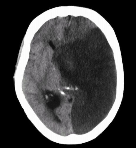
A 72 year-old man developed the abrupt onset of a global aphasia, right hemiplegia, left gaze preference and a right hemianopsia. Two days later, he lapsed into a coma and displayed decerebrate posturing. |

![]()
![]()
| Midline Shift and Herniation:
Axial
CT scan of the brain at the level of the basal ganglia. Note
the large infarction in the distribution of the left middle cerebral and
posterior cerebral arteries. There is significant midline shift secondary to the infarction
and associated edema, causing compression of the lateral ventricles
and contralateral hemisphere. Also note the calcified pineal gland,
which is normally a midline structure. In this case, it is shifted to the right. Such a picture of massive midline shift
secondary to infarction denotes a grave
prognosis.
The displacement of brain structures described above illustrates the Monro-Kellie doctrine, which states that in an adult the cranial volume is a constant. The cranial contents consist primarily of brain, cerebrospinal fluid (CSF) and blood vessels. If a mass such as a hematoma, tumor or edema develops, these elements must shift to accommodate the mass. Since the cranial volume is a constant, part of the cranial contents will herniate through the tentorial incisura to make room for the mass. The opposite occurs in a patient with loss of brain mass, such as occurs after a stroke, wherein the CSF spaces often enlarge to fill the void. |
Revised
11/30/06
Copyrighted 2006. David C Preston