|
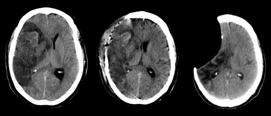
A 36 year-old man suffered a large right hemisphere ischemic stroke resulting in an acute left hemiplegia. Within a day, he was stuporous. To save his life and prevent herniation, a right sided craniectomy was performed. Two years later, the patient had still not returned to have the bone flap replaced. |
![]()
![]()
![]()
![]()
| "Missing" Brain s/p Craniectomy:
Axial CT scans. (Left) Day 3 after the onset on the stroke; (Middle)
Day 4 after the onset of the stroke, s/p right sided hemi-craniectomy; (Right)
Follow-up CT scan 2 years later. In the first scan (on the left), note the acute, massive right middle cerebral artery territory stroke associated with midline shift and impending herniation. In order to save the patient's life and prevent herniation, a right sided hemi-craniectomy was performed; note a large piece of skull has been removed to allow the brain to swell. Thus, the brain can then herniate through the craniectomy defect as opposed to herniating downward which would most likely be fatal. Also note the dark areas that indicate air in the craniectomy wound site following surgery (middle scan). In this case, the patient never returned to have the skull flap reimplanted. When he returned two years later for follow-up, there was a large defect in the right scalp. On the CT scan on the right, note the large area of missing brain rimmed by a thin layer of scalp tissue. This is an area of encephalomalacia, a result of the prior stroke. |
Revised
11/18/06.
Copyrighted 2006. David C Preston