|
A 27 year-old woman presented with progressive headaches and a focal seizure affecting the left side. |
![]()
![]()
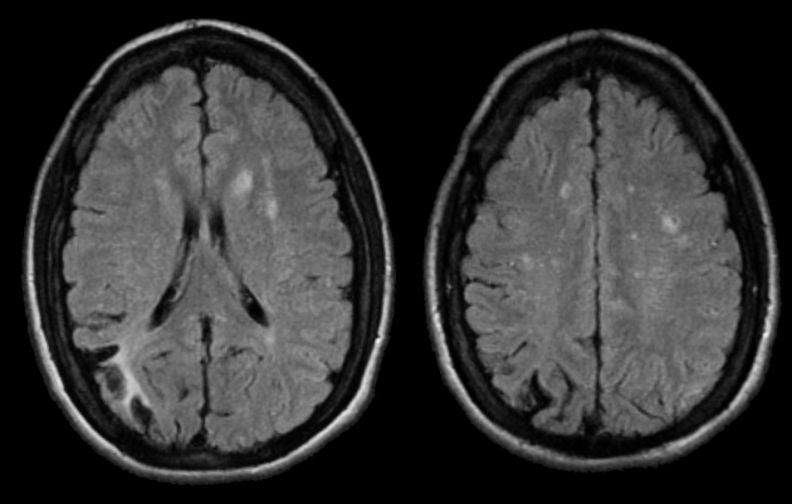
| Moyamoya Disease:
Flair Axial MRIs. Note the area of
encephalomalacia in the right posterior parietal cortex,
representing a remote stroke. On Flair MRI, fluid is
dark; thus, part of this old stroke is truly cystic due to missing,
necrotic brain. This remote stroke is in the distribution of the posterior
branches of the superior division of the middle cerebral artery. In
addition, note that there are several areas of small vessel strokes in
the deep white matter bilaterally. This picture is consistent
with both large and small vessel disease.
This patient has Moyamoya disease, a rare idiopathic disorder characterized by progressive narrowing of the distal internal carotid arteries and their branches. It is typically seen in children, although rarely reported in adults. As the carotid artery is compromised, there is progressive enlargement of the collateral circulation, especially among the lenticulostriate vessels. This results in a characteristic angiographic picture of a blush or "puff of smoke" in the area of the lenticulostriate vessels. Moyamoya disease typically presents with recurrent, progressive cerebral infarctions; in some cases, the fragile collateral vessels can rupture resulting in intracerebral hemorrhage. |
Revised
11/23/06
Copyrighted 2006. David C Preston