|
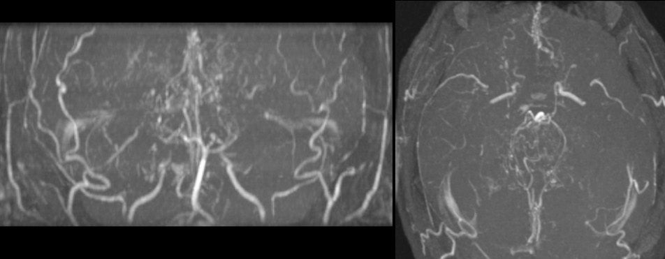
A 27 year-old woman presented with progressive headaches and a focal seizure affecting the left side. |
![]()
![]()
![]()
| Moyamoya Disease:
Intracranial MRAs; (Left) AP view;
(Right) Rostral-caudal projection. Note that these scans are extremely
abnormal (compare these scans to a normal
AP and
rostral-caudal intracranial MRA). The carotid arteries end
without the middle cerebral or anterior cerebral arteries being seen.
Although the basilar artery is present, the posterior cerebral arteries (PCAs)
terminate shortly after their origin. In addition, there are
prominent collaterals near the top of the carotid arteries which are a
characteristic finding in Moyamoya disease. This patient has Moyamoya disease, a rare idiopathic disorder characterized by progressive narrowing of the distal internal carotid arteries and their branches. It is typically seen in children, although rarely reported in adults. As the carotid artery is compromised, there is progressive enlargement of the collateral circulation, especially among the lenticulostriate vessels. This results in a characteristic angiographic picture of a blush or "puff of smoke" in the area of the lenticulostriate vessels. Moyamoya disease typically presents with recurrent, progressive cerebral infarctions; in some cases, the fragile collateral vessels can rupture resulting in intracerebral hemorrhage. |
Revised
11/23/06
Copyrighted 2006. David C Preston