|
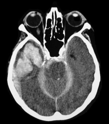
A 45 year-old woman was brought to the hospital in coma following a head injury sustained during a motor vehicle accident. |
![]()
![]()
![]()
| Multiple Types of Hemorrhage. Axial CT scan
without contrast. Note that there are three different types of hemorrhage in the same patient: subdural hematoma, intraparenchymal hemorrhage
(from contusion), and subarachnoid
blood. Subdural hematomas are recognized by their crescent
shape overlying and compressing the brain. They are arbitrarily
divided into three types: acute (< 4 days), subacute (4-21 days) and
chronic (> 21 days). Traumatic contusions consist of hemorrhage
and surrounding edema. The frontal poles and the temporal lobe tip
are the most common locations for cerebral contusions following head
injury, wherein the brain continues to move forward, striking the
inner skull, after the head has stopped moving. Subarachnoid
hemorrhage is the extravasation of blood into the subarachnoid space between the pial
and arachnoid membranes, in this case due to trauma.
In the acute stage, blood is bright on CT. Eventually in the chronic state, the blood turns dark. In the subacute stage, a variety of patterns can be seen. |
Revised
11/29/06.
Copyrighted 2006. David C Preston