|
A 43 year-old woman presented with headaches, nausea and vomiting. Two hours after admission to the hospital, she abruptly became unresponsive. |
![]()
![]()
![]()
![]()
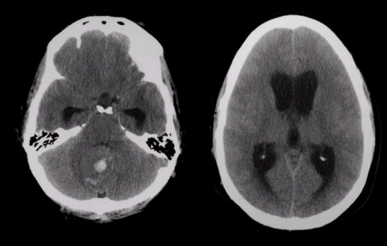
| Non-Communicating Hydrocephalus:
Axial CT scans. Note that there is acute blood in the vermis of the cerebellum. Looking closely,
one sees that the
cerebellum is enlarged by a mass which compresses the fourth ventricle. This is
an acute bleed into a tumor, compressing the fourth ventricle,
which resulted in acute non-communicating hydrocephalus.
Note also the dilated temporal horns. On the higher image (right
image), note the mildly enlarged lateral and third ventricles as well as the complete absence of sulci over the convexities. Surgical decompression of the cerebellar mass showed a malignant ependymoma. Hydrocephalus is recognized as enlarged ventricles out of proportion to the amount of cerebral atrophy. Non-communicating (obstructive) hydrocephalus occurs when the ventricular system is not in continuity with the subarachnoid space. Most often, the site of the blockage in non-communicating hydrocephalus is at the cerebral aqueduct, but rarely can occur at the foramen of Monro, the third ventricle, or the outlet of the fourth ventricle. Acute non-compensated, non-communicating (obstructive) hydrocephalus is a neurosurgical emergency as the non-compensated hydrocephalus results in a progressive increase in intracranial pressure, which if left unchecked will result in herniation and brain death. It is potentially treatable by shunting. |
Revised
11/29/06
Copyrighted 2006. David C Preston