|
A 34 year-old woman presented with focal seizures affecting the left face and arm. |
![]()
![]()
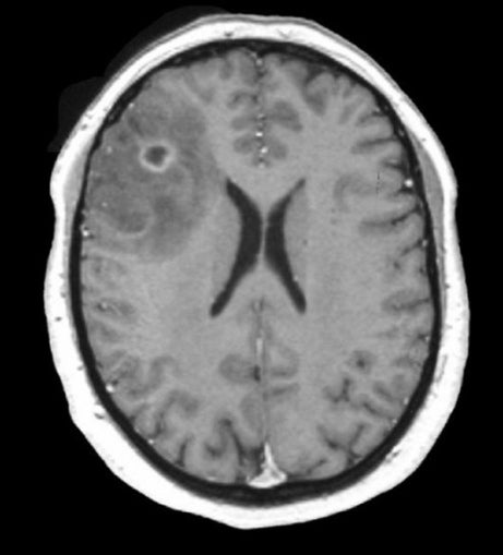
| Low Grade Glioma (Oligodendroglioma). T1-weighted with gadolinium
axial MRI. Note the well circumscribed lesion which enhances, with surrounding edema. Biopsy
demonstrated that the lesion was an oligodendroglioma.
Low-grade gliomas make up 15% of all primary intracranial brain tumors, and usually occur in young adults. They are named according to the specific type of glial cell that they derive from, and include astrocytomas, oligodendrogliomas, ependymomas, and mixed gliomas such as oligoastrocytomas that contain a mixture of different types of glial cells. Although biopsy is required to make a definitive diagnosis, the lack of contrast enhancement favors the diagnosis of a low grade glioma rather than a glioblastoma. Certain genetic conditions are predisposed to the formation of gliomas (e.g., neurofibromatosis, tuberous sclerosis). Clinical signs and symptoms depend on location. Headache and focal (or focal to generalized) seizures are common. Focal neurological deficits can occur. The World Health Organization scheme for grading gliomas from benign to progressively more malignant is as follows: ● Grade I - Pilocytic Pilocytic tumors are very benign histologically and typically occur in children. Low-grade tumors can be slow growing and controlled by surgical resection. If they recur, they are usually higher grade tumors. Anaplastic tumors are malignant tumors with mitoses and nuclear atypia. |
Revised
11/29/06.
Copyrighted 2006. David C Preston