|
A 66 year-old woman developed the abrupt onset of difficulty seeing on the left side, associated with numbness of the left side of the body. |
![]()
![]()
![]()
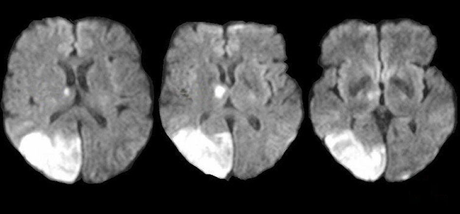
| Posterior Cerebral Artery (PCA) Infarction: Diffusion-weighted
MRIs. Note the bright signal in the
distribution of the right posterior cerebral artery. If one looks closely,
one can also see an infarction in the right thalamus.
This is the picture of an embolus to the proximal posterior cerebral artery.
The small perforating arteries to the thalamus arise from the proximal
posterior cerebral artery. Also note the subtle acute infarction in the left
occipital pole. This finding indicates involvement of the left posterior
cerebral artery as well. Putting all of these lesions together, the embolus was
likely situated at the top of the basilar artery occluding both posterior
cerebral arteries, and then broke up and propagated
into the right posterior cerebral artery proximally. The basilar artery bifurcates into two PCAs at its termination, which then course around the midbrain in the ambient and quadrigeminal cisterns to supply the medial / inferior temporal lobe and the medial occipital lobe. Small perforators arise from the proximal PCAs, which supply the cerebral peduncle as well as the thalamus (the latter known as thalamoperforators). Infarctions in the territory of the PCA most often result in a contralateral hemianopsia. Other signs and symptoms depend on whether the infarction involves the thalamoperforator territory, and whether the infarction is left vs. right sided. Proximal PCA involvement results in coexistent infarction of the lateral thalamus, with contralateral hemisensory findings. Infarction of either PCA may result in impaired memory (verbal on the left; spatial on the right). In addition, left PCA lesions are sometimes associated with alexia without agraphia (patients can write, but cannot read what they have written); transcortical sensory aphasia (a receptive aphasia similar to Wernicke's aphasia except that repetition is relatively spared); and Gerstmann's syndrome (a combination of acalculia, finger agnosia, agraphia, and right/left confusion). With right sided PCA lesions, patients may have a visual neglect. They may also have prosopagnosia, an inability to recognize familiar faces. |
Revised
11/23/06
Copyrighted 2006. David C Preston