|
A 21 year-old woman had an MRI done for complaints of intermittent headache. |
![]()
![]()
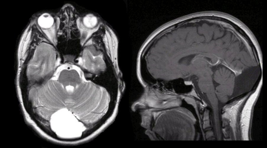
| Arachnoid Cyst of the Posterior Cranial Fossa:
(Left) T1-weighted axial MRI; (Right) T2-weighted axial MRI. Note
the large cystic lesion in the posterior fossa.
As the cyst contains fluid, it is dark on T1- and bright on
T2-weighted images. Arachnoid cysts are common congenital malformations. They contain fluid but generally do not communicate with the ventricular system. The most common locations are the Sylvian fissure and middle cranial fossa, suprasellar cistern, quadrigeminal cistern, cerebellopontine angle, posterior infratentorial midline, cerebral convexity and interhemispheric fissure. Most are asymptomatic and found incidentally on brain imaging studies. Rarely, they can enlarge and present as a mass lesion with focal neurological signs, seizures or hydrocephalus. |
Revised
11/29/06
Copyrighted 2006. David C Preston