|
A 59 year-old woman presented with an unstable gait. On exam, both legs were spastic with increased reflexes. |
![]()
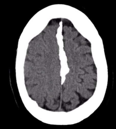
| Meningioma (Parafalcine). Axial CT scan. Note that the lesion
is calcified and arises from the falx in the midline. This is a common location
for a meningioma. In this case, the paracentral location resulted in the bilateral lower
extremity upper motor neuron signs. Meningiomas are common tumors that arise from the meninges and can occur within the spinal canal as well as intracranially. They are typically benign histologically, and can be surgically resected if they are in an accessible location. They are more common in women than men, and account for approximately 20% of all primary brain tumors. Similar to low grade gliomas, they grow very slowly. They may result in seizures, focal neurological signs, or both, depending on their location. Some of the more common locations for meningiomas include: ● Parasagittal (attached to the falx) |
Revised
11/29/06.
Copyrighted 2006. David C Preston