|
A 10 year-old girl presented with headaches and vomiting. |
![]()
![]()
![]()
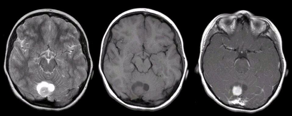
| Pilocytic Astrocytoma. (Left) T2-weighted
axial MRI; (Middle) T1-weighted axial MRI;
(Right) T1-weighted with gadolinium (Gad) axial MRI. Note the cystic mass in the region of the
midline of the cerebellum. There is an enhancing nodule surrounded by cystic fluid.
Surgical resection demonstrated the lesion to be a pilocytic
astrocytoma. The World Health Organization scheme for grading gliomas from benign to progressively more malignant is as follows: ● Grade I - Pilocytic Note that pilocytic tumors are very benign histologically. They are the most common astrocytic tumors in children and most often are located in the cerebellum. On imaging, they are cystic, often with an enhancing mural nodule. Because they most often occur in the cerebellum, initial symptoms tend to be headache and gait ataxia. Non-communicating hydrocephalus can occur. These lesions are typically treated by surgical resection and have an excellent prognosis. |
Revised
11/27/06.
Copyrighted 2006. David C Preston