|
A 62 year-old woman fell down the stairs and hit her head. In the ER, she was drowsy and mute. A head CT scan showed a large left sided subdural hematoma. She went for immediate surgical decompression. |
![]()
![]()
![]()
![]()
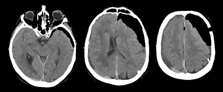
| Pneumocephalus: Axial CT scans - Post-operative day 1. Note the extremely dark areas, which are intracranial air. Also note the residual compression on the left lateral ventricle and midline shift. If one looks closely, one can also see a small right posterior, acute on chronic subdural hematoma (SDH). In this case, the intracranial air was introduced via the burr hole at the time of surgical decompression of the large left SDH. |
Revised
11/15/06.
Copyrighted 2006. David C Preston