|
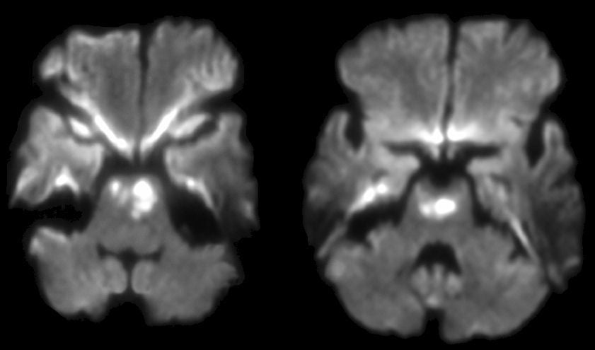
A 68 year-old woman was admitted with complaints of nausea, vomiting and right sided weakness. Within four hours, she was quadriplegic and could not speak. However, she could look up on command. An emergency angiogram demonstrated complete occlusion of the basilar artery. |
![]()
| Pontine and Midbrain Infarction: Diffusion-weighted axial
MRIs at the level of the pons (left) and upper pons/midbrain (right)
on Day 1. Note the bright signal within the base of the pons
and lower midbrain. This signal represents an acute ischemic injury. Note that the bright, symmetric signal seen in the frontal and temporal
lobes is a common diffusion artifact. Occlusion of the basilar artery may result in a "locked-in" syndrome, wherein the patient is completely quadriplegic and unable to speak. Infarction of the base of the pons interrupts the bilateral corticobulbar and corticospinal tracts, leaving the patient essentially disconnected from their body. Although such patients may be mistaken for being in a coma, they are actually awake and alert as the tegmentum is spared, the region where the ascending reticular activating system is located. Because control of vertical eye movements is located in the rostral midbrain, often the only voluntary movement is vertical gaze. |
Revised
11/29/06
Copyrighted 2006. David C Preston