|
An 82 year-old woman on coumadin for atrial fibrillation slipped and fell in the bathroom. After the fall, she had difficulty walking, with pain and bruising in her buttocks and down the back of her left leg. On admission, her INR was 6. Examination showed weakness of left knee extension and hip flexion, possibly limited by pain. Her patellar reflex was absent on the left and 2+ on the right, with normal ankle jerks bilaterally. She had decreased sensation over the medial aspect of her left lower leg. |
![]()
![]()
![]()
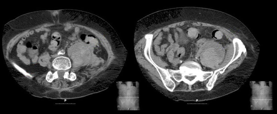
| Lumbar Plexopathy:
Axial CT scans of the abdomen and
pelvis. Note the large pelvic mass on the left. This is a
retroperitoneal hematoma within the substance of the psoas muscle. Note the
normal psoas muscle on the right.
Spontaneous retroperitoneal hemorrhage into the psoas muscle is often associated with coagulopathies (e.g., excessive anticoagulation, hemophilia). The hematoma places pressure on the underlying lumbar plexus, resulting in weakness of hip flexion (iliopsoas), knee extension (quadriceps) and hip adduction (thigh adductors). The knee reflex is typically depressed. Sensory loss may be present over the anterior thigh, medial thigh, and/or medial calf. Pain often interferes with assessment of muscle strength. |
Revised
11/29/06
Copyrighted 2006. David C Preston