|
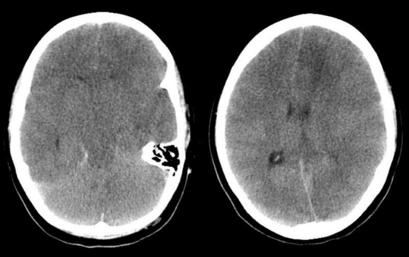
An 18 year-old woman presented with headaches followed by lethargy and then coma. On examination, she had papilledema. She withdrew the left side more than the right, to a painful stimulus. |
![]()
![]()
![]()
![]()
| Cerebral Venous Thrombosis. Axial CT scans. Note the diffuse cerebral edema manifested by effacement of the ventricles, cisterns and sulci, as well as the lack of differentiation between the gray and white matter. In addition, note the mass effect of the left hemisphere on the right (better seen on the image to the right). This is the CT scan appearance of impending herniation. The etiology cannot be determined from this scan alone. |
Revised
11/12/06.
Copyrighted 2006. David C Preston