|
A 72 year-old woman presented with progressive weakness of the left arm and leg. There was no facial weakness. On examination, she had increased tone and reflexes on the left side with a left Babinski sign. |
![]()
![]()
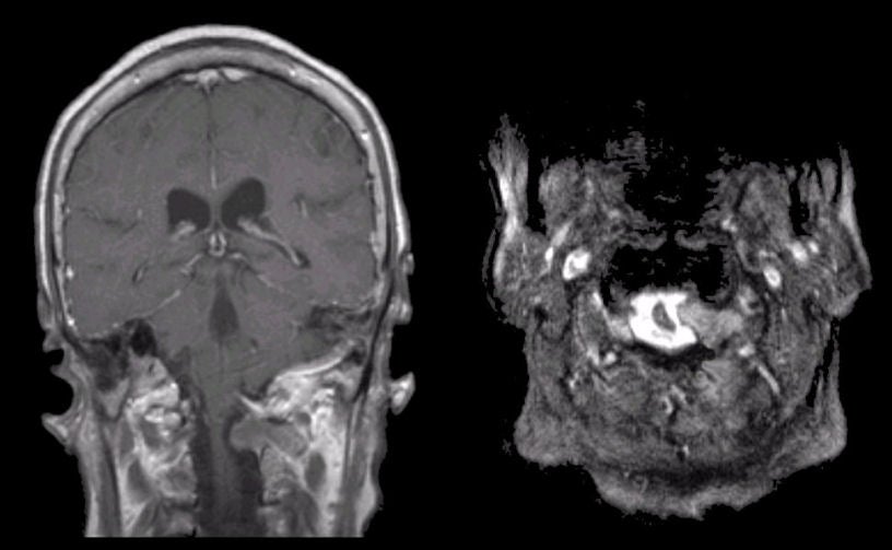
| Schwannoma:
(Left) T1-weighted with gadolinium coronal MRI; (Right)
T2-weighted axial MRI at the level of C1. Note the large somewhat dumb-bell shaped mass at the
C1 level. Also note the deformity of the adjacent spinal cord.
The mass is growing through the intervertebral foramen resulting in the
dumb-bell shape. At surgery, pathological
examination revealed a schwannoma.
Schwannomas are histologically benign tumors seen along the course of peripheral nerves, nerve roots, and cranial nerves
[especially cranial nerve V (trigeminal) and VIII (vestibulocochlear)].
They may occur in isolation or in association with
neurofibromatosis. They arise from the Schwann cells that
create the myelin sheath around peripheral nerves.
They result in symptoms when they disrupt the function of the nerve from
which they arise, or cause mass effect on adjacent structures. In
this case, symptoms resulted from compression of the high cervical
spinal cord when the Schwannoma grew along the C1 nerve root through
the intervertebral foramen. |
Revised
11/29/06
Copyrighted 2006. David C Preston