|
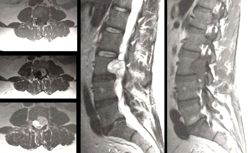
A 32 year-old man presented with progressive numbness and pain radiating from the back to the anterior and medial thigh. |
![]()
![]()
![]()
| Schwannoma:
MRI Scans of the Lumbar Spine; (Left Top) T1-weighted axial;
(Left Middle) T1-weighted with gadolinium axial; (Left Lower) T2-weighted axial; (Middle)
T2-weighted mid-sagittal; (Right) T1-weighted parasagittal MRI. Note the large nodular mass at the L3 level. On the
axial scans, the lesion enhances with gadolinium. On the
left lower image, one can clearly see that this lesion is extradural. On the
parasagittal scan on the right, note the normal nerve roots with surrounding
epidural fat, contrasted with the lesion that is growing through
and completely obliterating the L3 foramen. At surgery, pathological
examination revealed a schwannoma. Schwannomas are histologically
benign tumors seen along the course of peripheral nerves, nerve
roots, and cranial nerves [especially cranial nerve V (trigeminal)
and VIII (vestibulocochlear)]. They may occur in isolation or in
association with neurofibromatosis. They arise from the Schwann
cells that create the myelin sheath around peripheral nerves. They
result in symptoms when they disrupt the function of the nerve from
which they arise, or cause mass effect on adjacent structures. In
this case, symptoms resulted from compression of the cauda equina
when the Schwannoma grew along the L3 nerve root through the
intervertebral foramen. |
Revised
11/29/06
Copyrighted 2006. David C Preston