|
A 37 year-old man presented with numbness and pain on the left side of his face. |
![]()
![]()
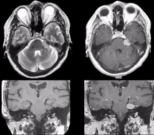
| Schwannoma:
(Top Left) T2-weighted axial MRI; (Top Right)
T1-weighted with gadolinium axial MRI; (Bottom Left) T1-weighted coronal MRI; (Bottom
Right)
T1-weighted with gadolinium coronal MRI. Note the nodular mass
adjacent to the pons
that enhances with gadolinium (right images). Surgical excision showed
that the mass was a schwannoma.
Schwannomas are histologically benign tumors seen along the course
of peripheral nerves, nerve roots, and cranial nerves [especially
cranial nerves V (trigeminal) and VIII (vestibulocochlear)]. They may
occur in isolation or in association with neurofibromatosis. They
arise from the Schwann cells that create the myelin sheath around
peripheral nerves. They result in symptoms when they disrupt the
function of the nerve from which they arise, or cause mass effect on
adjacent structures. |
Revised
11/29/06.
Copyrighted 2006. David C Preston