|
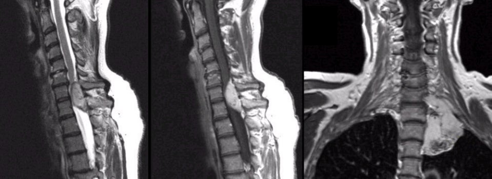
A 46 year-old woman presented with pain in the neck for several months followed by a rapidly progressive quadriparesis over two days. |
![]()
![]()
| Schwannoma:
(Left) T2-weighted sagittal MRI of the cervical spine;
(Middle) T1-weighted with gadolinium sagittal MRI of the cervical spine; (Right)
T1-weighted with gadolinium coronal MRI of the neck and lung. Note the extradural enhancing mass at the C7-C8 levels which compresses the spinal
cord. On the coronal scan, one can see that the mass extends down from the neck and invades the upper
lung. Excisional biopsy demonstrated a schwannoma. Schwannomas are histologically benign tumors seen along the course of peripheral nerves, nerve roots, and cranial nerves [especially cranial nerves V (trigeminal) and VIII (vestibulocochlear)]. They may occur in isolation or in association with neurofibromatosis. They arise from the Schwann cells that create the myelin sheath around peripheral nerves. They result in symptoms when they disrupt the function of the nerve from which they arise, or cause mass effect on adjacent structures. In this case, symptoms resulted from compression of the cervical spinal cord when the Schwannoma grew along the nerve root through the intervertebral foramen. |
Revised
11/28/06
Copyrighted 2006. David C Preston