|
A 72 year-old man presented with an unsteady gait. His neurological exam showed increased tone and hyperactive reflexes in the legs, with bilateral Babinski signs. Vibration sense was markedly decreased in the legs. |
![]()
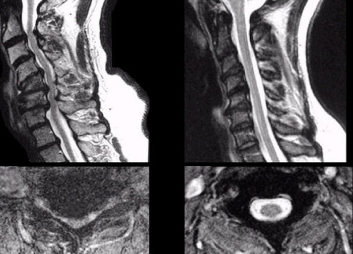
| Cervical Spondylosis:
T2-weighted MRI scans of the cervical spine;
(Left) Patient scans; (Right) Age-matched normal scans. Top
scans - Sagittal images; Bottom scans - Axial images. On T2-weighted
scans, CSF is white. In the patient scans on the left, note the severe stenosis
from C2-3 through C6-7.
The CSF is
nearly completely obliterated in the areas of compression. Also note
the marked deformity of the spinal cord on the axial image.
Cervical stenosis is a common cause of gait disturbance in the elderly. It results from progressive narrowing of the spinal column due to a combination of disk degeneration, bony spurs and ligament hypertrophy. |
Revised
11/29/06
Copyrighted 2006. David C Preston