|
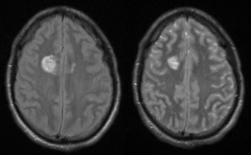
A 32 year-old woman presented with the abrupt onset of a severe headache associated with a mild left hemiplegia. |
![]()
![]()
| Subacute Late Intracerebral Hemorrhage: (Left) T1-weighted
axial MRI; (Right) T2-weighted axial MRI. Note
on the T1-weighted MRI, there is an area of hyperintense signal in the right posterior frontal
lobe. The same
area
is also hyperintense
on the T2-weighted MRI. This is the characteristic picture of subacute late hemorrhage (7-14 days old) on MRI. The hyperintense signal seen on both T1- and T2-weighted MRI is characteristic of extracellular methemoglobin. In this case, the hemorrhage was due to a deep arteriovenous malformation that bled. The characteristic findings of blood on MRI at different stages of timing are complex. To learn more, review the powerpoint slide show, Blood on MRI: Time-dependent Changes. |
Revised
11/11/06.
Copyrighted 2006. David C Preston.