|
A previously healthy 27 year-old woman developed a sudden, severe headache followed by nausea and vomiting. By the time she reached the hospital, she was poorly responsive. Her neurological examination was non-focal. |
![]()
![]()
![]()
![]()
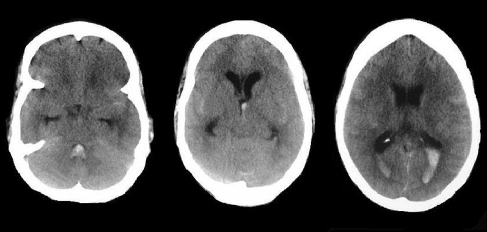
| Subarachnoid Hemorrhage.
Axial CT
scans without contrast. Note the bright areas which
signify blood in the fourth ventricle (left scan), the Sylvian fissure (middle scan)
and the posterior horns of the lateral ventricles (right scan). Also note the
enlarged ventricles (dilatation of the temporal horns) and effacement of the sulci over the convexity which indicate
increased intracranial pressure.
Acute hydrocephalus is a potential complication of subarachnoid
hemorrhage, either as a consequence of impaired CSF absorption or
obstruction of flow within the ventricular system. Subarachnoid hemorrhage (SAH) is the extravasation of blood into the subarachnoid space between the pial and arachnoid membranes. The most common causes of spontaneous SAH are rupture of a saccular (berry) aneurysm (80%) and rupture of an arteriovenous malformation (AVM) (10%). Aneurysm formation is also seen in the setting of mycotic aneurysms, as well as in association with some congenital disorders, including coarctation of the aorta, Marfan's syndrome, Ehlers-Danlos syndrome, fibromuscular dysplasia, and polycystic kidney disease. Causes of non-aneurysmal SAH include amyloid angiopathy, blood dyscrasias, fibromuscular dysplasia, Moyamoya disease, and vasculitis (10%). Aneurysms are usually located in the intracranial arteries which lack an external elastic lamina and have a very thin adventitia. They lie unsupported in the subarachnoid space. The early precursors of aneurysms are small outpouchings through defects in the media of the arteries. These defects expand as a result of hydrostatic pressure from pulsatile blood flow and blood turbulence. The probability of rupture is related to the tension on the aneurysm wall. From the Law of La Place, the tension on the wall is proportional to the diameter. Thus, the rate of rupture is directly related to the size of the aneurysm.. Aneurysms usually occur at arterial bifurcations and mostly arise from the anterior circulation of the Circle of Willis (85%). The most common sites of aneurysms include: • Posterior communicating artery Aneurysms can present with a variety of symptoms and signs, the following being the most common: • A sudden onset of severe headache
("thunderclap headache"), often described as the “worst headache of
my life" |
Revised
11/29/06.
Copyrighted 2006. David C Preston