|
A 65 year-old man with atrial fibrillation had the acute onset of left hemiplegia and a right gaze preference. Initial diffusion-weighted MRI scan indicated a large right MCA infarct. Two days later, the patient became lethargic. |
![]()
![]()
![]()
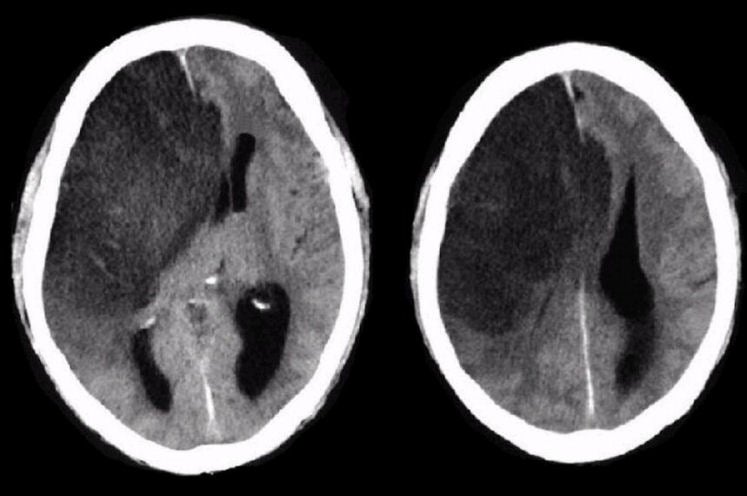
| Subfalcine Herniation: Axial CT scans: Note the large well defined infarction in the distribution of the proximal right MCA. Also note the herniation of the right cingulate gyrus under the falx (the fold of dura that lies between the cerebral hemispheres), which then compresses the lateral ventricles and left frontal lobe. Thus the term "subfalcine herniation". This is an ominous sign, denoting significant mass effect and increased intracranial pressure. Also note the amount of midline shift of the right hemisphere onto the left hemisphere. One useful way of measuring midline shift is examining how much the pineal gland has shifted off the midline. The pineal gland is normally a midline structure, and calcified in most adults, allowing it to be easily seen on CT imaging. |
Revised
11/29/06
Copyrighted 2006. David C Preston