|
A 60 year-old man presented with difficulty with his gait and bladder symptoms. |
![]()
![]()
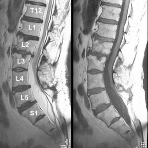
| Tethered Cord: (Left) T2-weighted sagittal MRI; (Right) T1-weighted
sagittal MRI. Note that the spinal cord ends at the L4-L5 level. This is
a tethered cord.
The spinal cord in adults normally terminates at the caudal level of the L1 vertebral body. A tethered spinal cord occurs when the cord becomes abnormally attached to tissue within the spinal canal, rather than floating free. This results in abnormal stretching of the spinal cord below the L1 vertebral body over time. It is more common in certain congenital disorders including myelomeningocele, lipoma or lipomyelomeningocele, diastematomyelia, dermal sinus tract, or tight filum terminale. The typical presentation is that of a slowly progressive myelopathy much like a syrinx, manifesting as a progressive gait abnormality (spastic or wide-based gait), weakness of the legs, sensory loss, and incontinence of bowel and bladder. Patients with symptomatic tethered cords often present with orthopedic deformities, including pes varus, valgus, or cavus; recurrent hip dislocations; or rotational abnormalities of an extremity or extremities. An isolated presentation of scoliosis is not uncommon. |
Revised
11/29/06
Copyrighted 2006. David C Preston