|
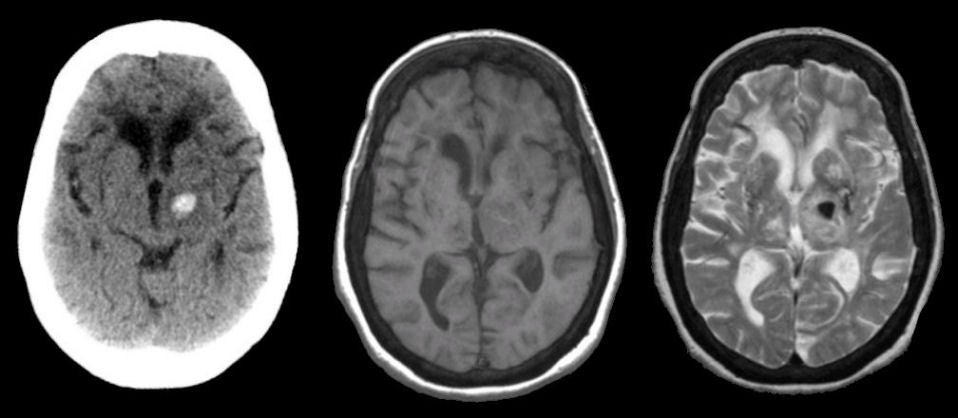
A 52 year-old hypertensive man developed a headache, nausea and vomiting, associated with right sided numbness that slowly worsened over 30 minutes. |
![]()
![]()
![]()
| Thalamic Intracerebral Hemorrhage: (Left) Axial CT scan; (Middle) T1-weighted axial MRI; (Right) T2-weighted axial MRI. On the CT scan (left image), note the bright signal in the left thalamus, indicating acute hemorrhage. On the T1-weighted image (middle scan), the abnormality is not well seen, as the signal is isointense. However, on the T2-weighted image (right scan), the signal is dark, representing deoxyhemoglobin. This is the MRI picture of an acute/subacute hemorrhage. |
Revised
11/23/06.
Copyrighted 2006. David C Preston