|
A 64 year-old hypertensive man developed a headache, nausea and vomiting, associated with right sided numbness. Within 3 hours, he lapsed into a coma. |
![]()
![]()
![]()
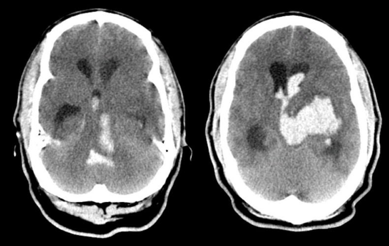
| Thalamic Intracerebral Hemorrhage with Ventricular Extension:
Axial CT scans. Note
that there is a large hemorrhage in the region of the left thalamus with
extension to the lateral, third and fourth ventricles. In addition, there is
associated hydrocephalus. This is the picture of impending brain herniation. This is one of the common sites of hypertensive intracerebral hemorrhage. Early neurological symptoms include contralateral sensory loss. With mass effect, patients develop headache, nausea and vomiting. As the lesion expands, patients may become lethargic due to direct compression on the upper brainstem structures or obstructive hydrocephalus (as seen above). Eye movement abnormalities, especially impaired vertical gaze, are common in thalamic mass lesions. |
Revised
11/23/06.
Copyrighted 2006. David C Preston