![]()
| Venous Malformation (Angioma):
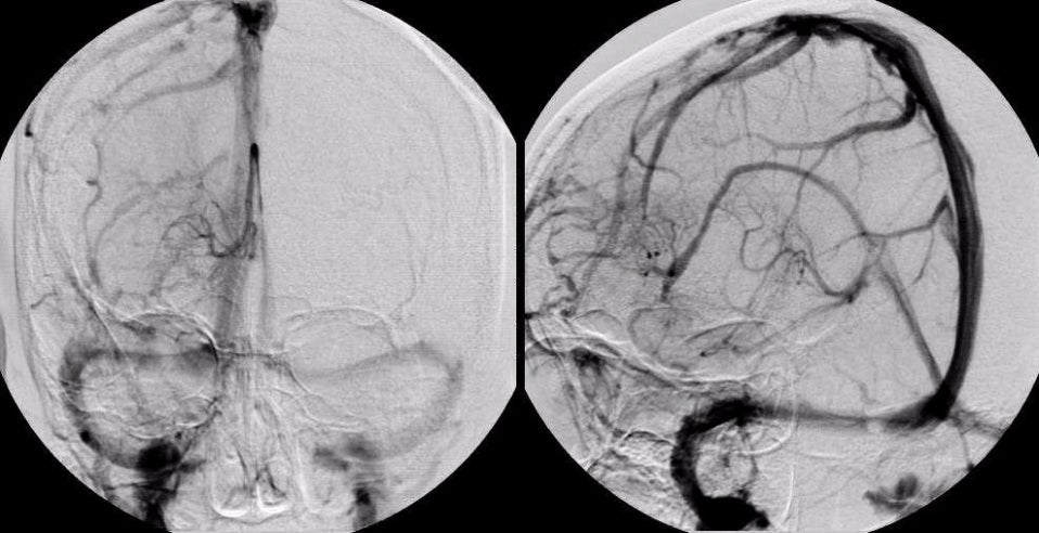
Cerebral angiogram, left carotid injection, venous phase; (Left) AP view; (Right) Oblique view. Note the abnormal venous structure
draining into the inferior sagittal sinus. This lesion is a venous
angioma. Venous angiomas are congenital malformations of the
cerebral veins, composed of several small veins that join to drain
into a larger venous trunk which then drains into a dural sinus.
On CT and MRI scans, venous angiomas enhance with contrast, and have
the appearance of a "medusa head". They are associated with other vascular malformations in about
15-30% of patients, most frequently cavernous angiomas. Most times, venous angiomas are incidental findings on brain imaging. However, with the advent of MRI and its high sensitivity in detecting remote hemorrhage, venous angiomas are more often recognized as a source of intracerebral hemorrhage. Thus, they are a potential etiology of seizures, focal neurological findings, or rarely symptomatic intracranial hemorrhage. |
Revised
11/29/06
Copyrighted 2006. David C Preston