|
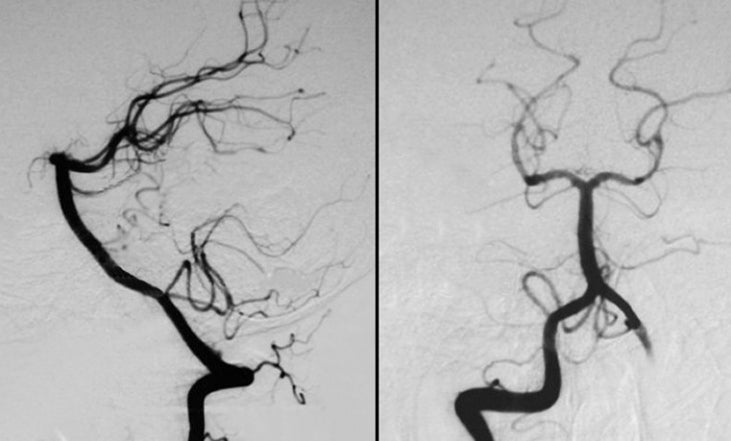
Vertebral Basilar
Angiogram. (Left) lateral view; (Right) AP view.
This study was performed by
injecting contrast into the right vertebra artery, which is well
opacified. There is also some retrograde flow of contrast down
the left vertebral artery which allows visualization of the distal
portion of the vertebral artery and posterior inferior cerebellar
artery PICA on that side.
The Posterior Cerebral Artery
(PCA) is commonly divided into four numbered segments: (1) the P1
segment runs from the top of the basilar to the origin of the
posterior communicating artery in the interpeduncular cistern; (2)
the P2 segment runs through the ambient cistern from the
origin from the posterior communicating artery around the cerebral
peduncle; (3) the P3 segment runs through the quadrigeminal
cistern to where the calcarine sulcus originates; and (4) the
P4 segments runs from the calcarine sulcus to over the cortex
|