|
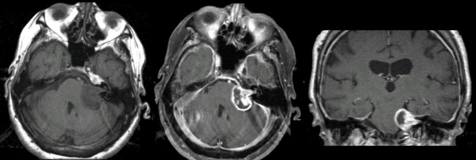
A 56 year-old woman presented with progressive headaches, loss of hearing in the left ear, and falling to the left. |
![]()
![]()
![]()
![]()
| Acoustic Neuroma:
(Left) T1-weighted axial MRI; (Middle) T1-weighted with
gadolinium axial MRI; (Right) T1-weighted with gadolinium coronal MRI. Note the
large nodular mass
adjacent to the lower pons on the left. The mass enhances with gadolinium. In
addition, note the compression of the middle cerebellar peduncle and
4th ventricle. On excision, this lesion was
a schwannoma of the vestibulocochlear nerve.
Schwannomas are histologically benign tumors seen along the course
of peripheral nerves, nerve roots, and cranial nerves [especially
cranial nerves V (trigeminal) and VIII (vestibulocochlear)]. They may
occur in isolation or in association with neurofibromatosis. They
arise from the Schwann cells that create the myelin sheath around
peripheral nerves. They result in symptoms when they disrupt the
function of the nerve from which they arise, or cause mass effect on
adjacent structures. |
Revised
11/29/06.
Copyrighted 2006. David C Preston