|
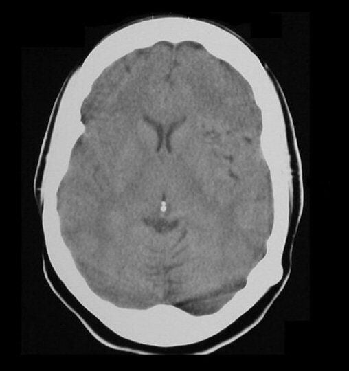
A 36 year-old woman presented with headaches and focal seizures affecting her language and the right side of her body. |
![]()
| Arteriovenous Malformation (AVM): Axial CT scan (level of
the basal ganglia). The scan initially appears normal. However, if
one looks closely, a collection of dark vascular spaces are seen in
the region of the left Sylvian fissure. Without contrast or an
angiogram, AVMs may be easily missed.
Arteriovenous malformations (AVMs) are a congenital abnormality of blood vessels. They consist of a tangle of abnormal vessels supplied by arterial feeders and often drained by large dilated veins. AVMs most often occur in isolation. Rarely, they are associated with genetic disorders, among them: Osler-Weber-Rendu syndrome (hereditary hemorrhagic telangiectasia), Sturge-Weber disease, and von Hippel-Lindau syndrome. AVMs are often asymptomatic. Symptoms, when present, may include: • headaches (in some cases a unilateral throbbing headache, mimicking a migraine headache) • seizures (focal, or focal to generalized) • focal neurological deficits • bleeding (may mimic subarachnoid hemorrhage from an aneurysm; bleeding from AVMs account for 2% of all strokes) Larger AVMs are often seen on CT or MRI. Angiography is required to define the vascular anatomy and plan appropriate treatment. Treatment may involve surgical resection, embolization or radiotherapy. |
Revised
11/29/06
Copyrighted 2006. David C Preston