![]()
![]()
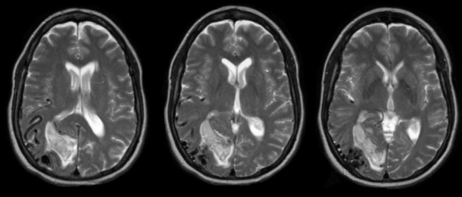
| Arteriovenous Malformation (AVM):
T2-weighted axial MRIs. Note the dark flow
voids in the right occipital lobe near the cortex. These
are the dilated vessels of the AVM. The bright signal in the occipital
cortex is a subacute intracerebral hemorrhage (intracellular methemoglobin
is bright on T2- and T1-weighted scans). Note how easy it is to
diagnose the AVM by MRI scan compared to the earlier CT scan. Arteriovenous malformations (AVMs) are a congenital abnormality of blood vessels. They consist of a tangle of abnormal vessels supplied by arterial feeders and often drained by large dilated veins. AVMs most often occur in isolation. Rarely, they are associated with genetic disorders, among them: Osler-Weber-Rendu syndrome (hereditary hemorrhagic telangiectasia), Sturge-Weber disease, and von Hippel-Lindau syndrome. AVMs are often asymptomatic. Symptoms, when present, may include: • headaches (in some cases a unilateral throbbing headache, mimicking a migraine headache) • seizures (focal, or focal to generalized) • focal neurological deficits • bleeding (may mimic subarachnoid hemorrhage from an aneurysm; bleeding from AVMs account for 2% of all strokes) Larger AVMs are often seen on CT or MRI. Angiography is required to define the vascular anatomy and plan appropriate treatment. Treatment may involve surgical resection, embolization or radiotherapy. |
Revised
11/29/06
Copyrighted 2006. David C Preston