![]()
![]()
![]()
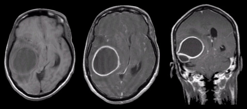
| Brain Abscess:
(Left) T1-weighted axial MRI; (Middle) T1-weighted with
gadolinium axial MRI; (Right) T1-weighted with gadolinium coronal MRI. Note the obvious
ring enhancement on the gadolinium enhanced scans. Also note the prominent
mass effect on the third ventricle and the lateral ventricles. Neurosurgical
aspiration revealed pus and numerous gram positive cocci. Intracranial abscesses can occur in the epidural and subdural space as well as in the brain parenchyma. Infection most often occurs from spread through the blood system, or from direct invasion of an infection from an adjacent structure (e.g., sinusitis, otitis, mastoiditis, etc). Patients most often present subacutely over days to a few weeks with fever, headache, and/or focal neurological signs, including seizures. |
Revised
11/23/06
Copyrighted 2006. David C Preston