|
A 17 year-old girl underwent an MRI scan as part of a headache evaluation. Her neurological examination was normal. |
![]()
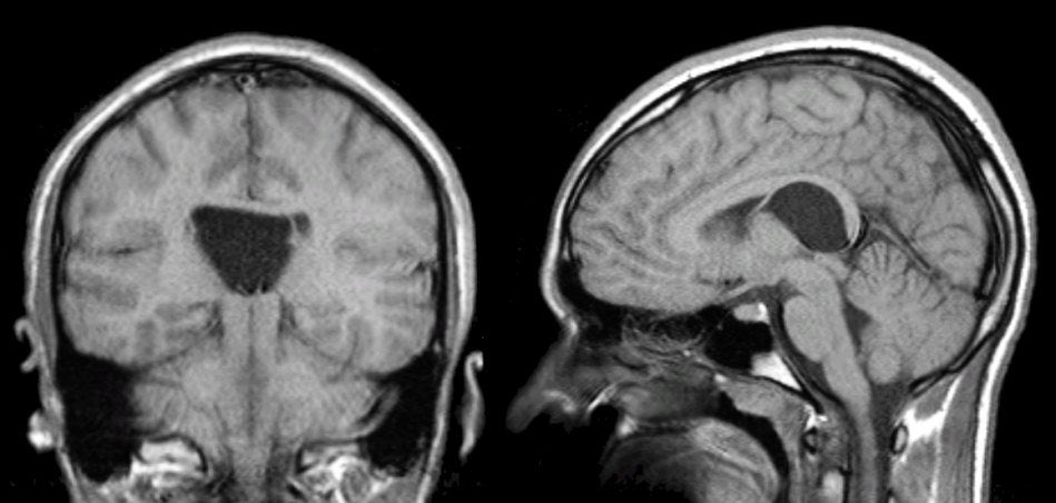
| Cavum Veli Interpositi: (Left) T1-weighted coronal MRI;
(Right) T1-weighted coronal MRI. Note the large triangular shaped
fluid filled structure in the midline. This is a Cavum Veli
Interpositi. In utero, three potential midline cavities may occur along ventricles. From anterior to posterior, these are the Cavum Septum Pellucidum, Cavum Vergae and Cavum Veli Interpositi, respectively. Most often, these cavities disappear between the seventh month in utero and the second year of life, but in some cases may persist. The Cavum Veli Interpositi is thought to be a fusion defect of the velum interpositum. The velum interpositum is a potential subarachnoid space in the tela choroidea that forms the roof of the third ventricle. The tela choroidea is composed of the fusion of pia and ependyma. It also contains the internal cerebral veins. The velum interpositum is located between the fornix and its attached choroid above, and the choroid forming the roof of the 3rd ventricle below. It is effectively an anterior extension of the quadrigeminal cistern, and recognized by its characteristic triangular shape. In individuals in whom this space is prominent, it is called a Cavum Veli Interpositi, and does not have any clinical significance. However, if it is associated with an arachnoid cyst, there may be mass effect on adjacent structures. It is often misidentified as the third ventricle. However, remember, the third ventricle is slit-shaped with parallel walls, whereas the velum interpositum is located in the roof of the third ventricle and is triangular in shape. |
Revised
11/29/06
Copyrighted 2006. David C Preston