|
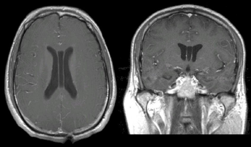
A 55 year-old man underwent an MRI scan as part of a syncope evaluation. His neurological examination was normal. |
![]()
| Cavum Vergae: (Left) T1-weighted with gadolinium axial
MRI; (Right) T1-weighted with gadolinium coronal MRI. Note the extra
CSF space between the two lateral ventricles. This is a Cavum
Vergae. Cavum Vergae is a normal anatomic variant, and is the
posterior extension of a Cavum Septum Pellucidum, another normal
anatomic variant. The cavity was first described by the Italian
anatomist, Andrea Verga, in 1851. It may exist as a separate cavity,
or may communicate with the Cavum Septum Pellucidum. Its incidence
is estimated at approximately 2%. It has no clinical significance.
It is sometimes termed the "6th ventricle," a misnomer, since it
does not contain cerebrospinal fluid and is not lined by ependyma.
In utero, three potential midline cavities may occur along ventricles. From anterior to posterior, these are the Cavum Septum Pellucidum, Cavum Vergae and Cavum Veli Interpositi, respectively. Most often, these cavities disappear between the seventh month in utero and the second year of life, but in some cases may persist. |
Revised
11/30/06
Copyrighted 2006. David C Preston