![]()
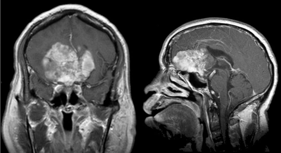
| Glioblastoma Multiforme (Frontal Lobe).
(Left) T1-weighted with gadolinium coronal MRI; (Right) T1-weighted with
gadolinium sagittal MRI. Note the large enhancing mass in the frontal lobes.
This pattern of a frontal tumor crossing over the midline to the
contralateral frontal lobe via the corpus callosum is known as a
"butterfly" pattern. Biopsy showed glioblastoma multiforme.
Glioblastoma multiforme (GBM), also referred to as a Grade IV
astrocytoma, is the most common type of primary brain tumor. It is a
malignant tumor that carries a very poor prognosis, and typically
results in death in 2 years. On CT and MRI imaging, the tumor is
often large, irregular and infiltrative, and located in the white
matter with surrounding edema. Histologically, the tumor is highly
cellular and anaplastic with necrosis. Associated hemorrhage is not
uncommon. Clinically, patients present with slowly progressive focal neurological signs, and signs of increased intracranial pressure (i.e., headache, nausea, and vomiting). Seizures may be an initial presentation or may occur later in the course. |
Revised
11/23/06.
Copyrighted 2006. David C Preston