|
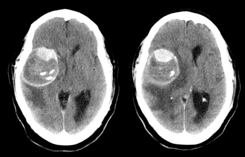
A 42 year-old woman developed headaches and slowly progressive left sided weakness over two months. |
![]()
![]()
![]()
| Middle Cerebral Artery Giant Aneurysm. Axial CT scans without contrast. Note the large mass in the right frontal/temporal lobes associated with mass effect and vasogenic edema. Note that the mass is hetereogenous: the very bright areas are calcifications whereas the slightly bright areas are clot within the aneurysm (see the MRI image). Also note the circular pattern of the mass. This is a giant aneurysm of the middle cerebral artery, which is partially thrombosed. Although most aneurysms present with bleeding, occasionally they enlarge and present with focal neurological findings from mass effect on adjacent structures. |
Revised
11/23/06.
Copyrighted 2006. David C Preston