|
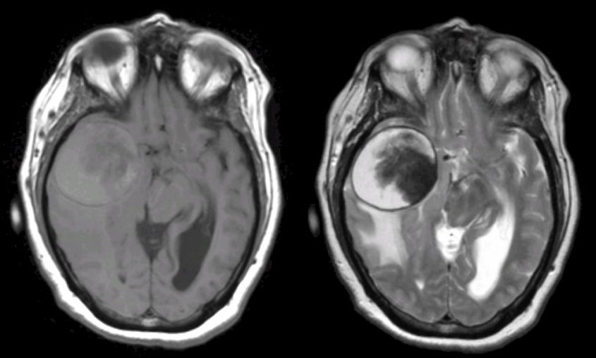
A 42 year-old woman developed headaches and slowly progressive left sided weakness over two months. |
![]()
![]()
![]()
| Middle Cerebral Artery Giant Aneurysm. Axial MRI scans; (Left) T1-weighted; (Right) T2-weighted. Note the large mass in the right frontal/temporal lobes associated with mass effect and vasogenic edema. On the T2-weighted scan, the mass has both white and dark signal, representing partially clotted blood (the dark signal is deoxyhemoglobin, whereas the bright signal is methemoglobin). This is a giant aneurysm of the middle cerebral artery which is mostly thrombosed. |
Revised
11/23/06.
Copyrighted 2006. David C Preston