![]()
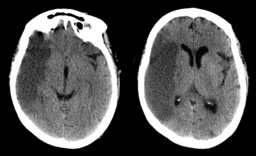
| Distal Stem Middle Cerebral Artery (MCA) Infarction:
Axial CT scans 3 days after presentation. Note the dark signal involving
the distribution of both the superior and inferior divisions of the
middle cerebral artery, but sparing the basal ganglia. This syndrome
is most commonly caused by an embolus that blocks the distal middle
cerebral artery stem, after the take-off of the lenticulostriate
vessels. The internal carotid artery terminates in a larger MCA and smaller anterior cerebral artery. The MCA runs horizontally to the Sylvian fissure, giving off the lenticulostriate vessels. These small perforating vessels supply the basal ganglia and internal capsule. The MCA then typically bifurcates into a superior and inferior division. The superior division supplies the lateral frontal and superior parietal lobes, whereas the inferior division predominantly supplies the lateral temporal and inferior parietal lobes. Occlusions of the distal stem of the MCA affect both the superior and inferior divisions, but spare the lenticulostriates. Complete infarctions from a distal MCA stem occlusion result in a contralateral hemiplegia (affecting the lower face and arm more than the leg); contralateral hemisensory loss with a similar distribution; and a contralateral visual field deficit. With an infarct in the dominant hemisphere, there is often an associated global aphasia (expressive and receptive); with a non-dominant infarct, there is often a neglect syndrome and impairment of visuospatial skills (e.g., drawing, copying, dressing). |
Revised
11/15/06
Copyrighted 2006. David C Preston